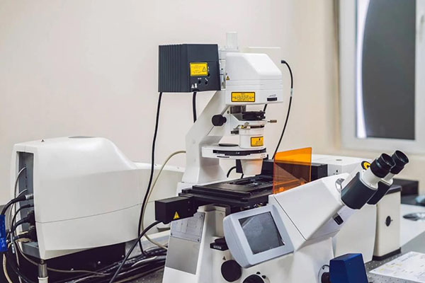
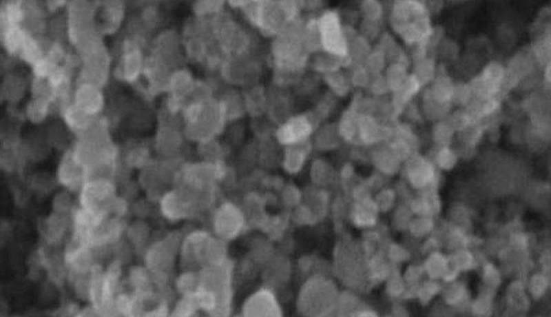
Scanning Electron Microscopy (SEM) is a powerful imaging technique that has revolutionized the way we visualize and understand the microstructure of various materials and biological specimens. SEM offers numerous advantages, such as high magnification, large depth of field, and straightforward sample preparation. In this blog, we will explore the key advantages of SEM and the factors that influence its resolution, magnification, depth of field, and contrast.
Advantages of Scanning Electron Microscopy (SEM)
High Magnification: SEM offers a wide range of magnification levels, typically ranging from 20 to 200,000 times. This capability allows researchers to investigate structures at the nanoscale and beyond, revealing intricate details not observable with other conventional microscopes.
Large Depth of Field: Unlike optical microscopes, SEM has a substantial depth of field, enabling clear visualization of 3D surface structures. This is especially valuable when studying rough or uneven surfaces of various samples.
Sample Preparation: Preparing samples for SEM analysis is relatively simple compared to other high-resolution microscopy techniques like transmission electron microscopy (TEM). Samples are coated with a thin conductive layer, making them suitable for SEM imaging without complex sample preparation procedures.
Factors Affecting SEM Resolution
Resolution is a critical aspect of SEM imaging and refers to the ability to distinguish fine details in the specimen. Several factors influence SEM resolution:
A. Electron Beam Spot Size: The minimum spot size of the incident electron beam is a fundamental factor affecting resolution. With advanced electron gun technologies, SEMs can achieve spot sizes as small as 3 nm with field emission electron guns.
B. Electron Beam Spread in the Sample: The degree of electron beam spread within the sample depends on the electron beam’s energy and the atomic number of the sample. Higher beam energies and lower atomic number samples lead to larger interaction volumes, reducing resolution.
C. Imaging Mode and Signal Modulation: Different imaging modes, such as secondary electron imaging and backscattered electron imaging, affect resolution differently. Secondary electrons, being low in energy, provide higher-resolution images compared to backscattered electrons, which have higher penetration depths and lateral spread.
Magnification in SEM
The magnification in SEM can be represented as M = Ac/As, where Ac is the length of the image on the fluorescent screen, and As is the scan amplitude of the electron beam on the sample surface. SEM allows magnifications ranging from 20 to 20,000 times, bridging the gap between optical microscopes and transmission electron microscopes.
Depth of Field in SEM
Depth of field refers to the range in front and behind the focal point where all object points form a sharp image with sufficient resolution. SEM provides a depth of field ten times greater than transmission electron microscopy and hundreds of times greater than optical microscopy. The depth of field depends on the critical resolution (d0) and the angle of incidence (αc) of the electron beam. Small apertures and large working distances result in a smaller incident angle, thereby increasing the depth of field.
Contrast in SEM
Contrast in SEM is achieved through surface morphology and atomic number contrast. Surface morphology contrast arises from the sample’s unevenness, while atomic number contrast relies on the sensitivity of backscattered electrons, absorbed electrons, and X-rays to variations in atomic numbers. Higher atomic numbers in the sample result in brighter images. Conversely, secondary electrons are less affected by atomic number differences, making them less suitable for high-resolution imaging of materials with small atomic number variations.
Conclusion
Scanning Electron Microscopy has proven to be an indispensable tool for researchers and scientists across various disciplines. Its high magnification, large depth of field, and straightforward sample preparation have unlocked new possibilities in the study of materials and biological specimens.
Understanding the factors influencing resolution, magnification, depth of field, and contrast in SEM helps researchers optimize imaging parameters and obtain precise and detailed results. With ongoing advancements in electron microscopy technology, SEM remains at the forefront of cutting-edge research and discovery.


